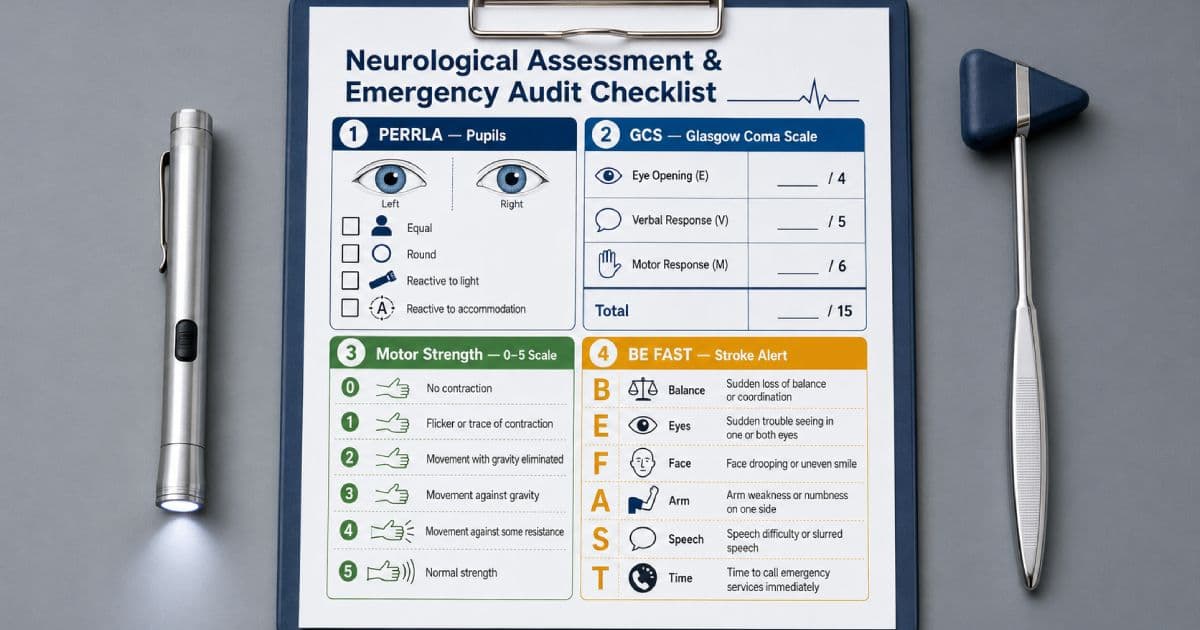
A neurological change may be subtle before it becomes catastrophic.
A patient may become slightly slower to answer, develop a mild arm drift, or show a new difference in pupil size before a dramatic decline occurs. These findings can be early signs of stroke, expanding intracranial bleeding, seizure activity, medication effects, hypoglycemia, or rising intracranial pressure (ICP).
The bedside nurse is often the first clinician to notice that something has changed.
A useful neuro assessment for nurses must therefore be structured, reproducible, and compared with the patient's established baseline. The goal is not simply to complete a checklist. The goal is to identify a meaningful change, protect the patient, and escalate care without delay.
This guide covers:
- Level of consciousness and orientation
- Speech and command following
- Pupil size, equality, shape, and reactivity
- Bilateral motor strength and pronator drift
- Glasgow Coma Scale scoring
- BE FAST stroke warning signs
- Decorticate and decerebrate posturing
- Early and late signs of increased ICP
- Clear neuro assessment documentation
What Is a Neurological Assessment?
A neurological assessment evaluates how well the central and peripheral nervous systems are functioning.
The depth of the assessment depends on the clinical situation.
A complete neurological examination may include:
- Mental status
- Cranial nerves
- Motor function
- Sensory function
- Coordination
- Balance and gait
- Reflexes
A focused bedside neuro check is shorter. It usually emphasizes the findings most likely to change quickly:
- Level of consciousness
- Orientation
- Speech
- Pupils
- Facial symmetry
- Extremity movement and strength
- Sensation
- Glasgow Coma Scale, when ordered or clinically appropriate
- Vital-sign patterns associated with deterioration
Neuro checks may be ordered after:
- Stroke or transient ischemic attack (TIA)
- Head injury
- Neurosurgery
- Seizure
- Intracranial hemorrhage
- Sedation or anesthesia
- A fall with possible head trauma
- Administration of thrombolytic therapy
- A sudden change in mental status
The prescribed frequency may range from routine assessments to checks every 15 minutes during a high-risk period. Follow the order, unit standard, and escalation policy.
The Most Important Rule: Compare With Baseline
One isolated finding rarely tells the whole story.
The same GCS score, pupil size, or strength grade can mean different things in different patients. A patient with a chronic right-sided weakness after an old stroke may be stable at 3/5 strength. Another patient whose right arm was 5/5 an hour ago and is now 3/5 may be experiencing an acute emergency.
Before beginning, review:
- The previous neurological assessment
- The patient's normal cognitive status
- Known residual deficits
- Recent sedatives, opioids, paralytics, or anesthetics
- Eye surgery, ocular trauma, or baseline anisocoria
- Language, hearing, and communication barriers
- Intubation, aphasia, or other barriers to GCS testing
- The time of the last normal assessment
Prepare for the Assessment
Before touching the patient:
- Perform hand hygiene.
- Confirm the patient's identity.
- Explain what you are assessing.
- Provide adequate lighting.
- Reduce unnecessary noise.
- Check whether glasses or hearing aids are normally used.
- Assess for immediate airway, breathing, and circulation threats.
- Review the most recent neuro findings and vital signs.
If the patient appears acutely different, obtain help early.
When stroke is possible, note the time and determine the last known well time while the emergency response is being activated. Do not allow a lengthy assessment to delay imaging or treatment.
A Step-by-Step Focused Neuro Assessment
Use the same sequence each time. Consistency makes trends easier to recognize.
Step 1: Observe Before You Stimulate
Watch the patient as you enter the room.
Ask:
- Are the eyes open spontaneously?
- Does the patient track movement?
- Is the patient speaking appropriately?
- Is breathing regular?
- Is the face symmetrical at rest?
- Are both sides of the body moving?
- Is the patient restless, unusually still, or difficult to arouse?
Spontaneous behavior is valuable. Immediately calling the patient's name or touching the patient can make it harder to determine whether eye opening was truly spontaneous.
Step 2: Assess Level of Consciousness
Level of consciousness (LOC) describes the patient's degree of wakefulness and ability to respond.
A change in LOC is one of the most sensitive indicators of neurological deterioration. It may occur before changes in pupils or vital signs.
Step 3: Assess Orientation and Speech
If the patient is awake, assess orientation and listen to how the patient communicates.
Step 4: Assess Pupils
Measure pupil size, compare both pupils, inspect shape, and test the light response.
Step 5: Assess the Face and Extremities
Look for facial asymmetry, arm drift, unilateral weakness, abnormal movement, and sensory changes.
Step 6: Calculate the GCS When Indicated
Score eye opening, verbal response, and best motor response separately.
Step 7: Compare, Document, and Escalate
Compare the findings with baseline and the previous neuro check.
Report new or worsening findings immediately.
Level of Consciousness and Orientation
Do not use orientation and level of consciousness as interchangeable terms.
A patient may be awake but confused. Another patient may know their name when awakened but be unable to remain alert.
Document both wakefulness and orientation.
Progressive Levels of Consciousness
Terms vary slightly across organizations. Use your facility's approved definitions and describe the observable response whenever possible.
| Level | Typical bedside finding |
|---|---|
| Alert | Awake spontaneously, attentive, and responds appropriately |
| Lethargic or somnolent | Drowsy and may fall asleep, but awakens to voice and responds appropriately or with mild delay |
| Obtunded | Markedly reduced alertness; requires repeated verbal or tactile stimulation and gives limited responses |
| Stuporous | Responds only to vigorous or noxious stimulation and does not sustain interaction |
| Comatose | Cannot be aroused and shows no purposeful response to verbal or noxious stimulation |
Avoid charting only “sleepy” or “hard to wake.” These phrases are vague.
Document what you did and how the patient responded.
Patient opens eyes after repeated verbal prompting, states name, follows one-step command, then falls asleep within 10 seconds.That description is more reproducible than “patient lethargic.”
Assessing Orientation: A&Ox4
Assess four distinct domains.
Person
Ask:
“Can you tell me your full name?”
Do not ask, “Do you know who you are?” A yes-or-no question does not prove orientation.
Place
Ask:
“Can you tell me where you are right now?”
Accept a response appropriate to the situation. The patient may identify the hospital without knowing the building's exact name.
Time
Ask:
“What month and year is it?”
The exact date is more demanding and may be missed by otherwise oriented patients. Compare the response with baseline and clinical context.
Situation
Ask:
“Can you tell me why you are here?”
This evaluates understanding of the current circumstances.
Document the specific domains rather than only “A&Ox3” when possible.
Alert; oriented to person and place. States the year is 2024 and cannot explain reason for admission. Speech clear. Follows two-step commands.Assess Attention and Command Following
Orientation alone does not establish normal cognition.
Ask the patient to follow a simple command:
- “Open and close your eyes.”
- “Show me two fingers.”
- “Lift your right arm.”
Then consider a two-step command if appropriate:
“Touch your left ear with your right hand, then point to the ceiling.”
Inability to follow a command may reflect:
- Reduced consciousness
- Receptive aphasia
- Hearing impairment
- Language mismatch
- Inattention or neglect
- Pain or weakness
- Sedation
- Delirium
Do not assume the cause without assessing the barrier.
Assess Speech and Language
Listen for:
- Slurred articulation
- Incorrect or nonsensical words
- Difficulty naming familiar objects
- Trouble understanding instructions
- Delayed responses
- New hoarseness
- Inability to repeat a sentence
Dysarthria
Dysarthria is a motor speech problem. The patient knows what they want to say, but speech may sound slurred, slow, or poorly articulated.
Aphasia
Aphasia is an impairment of language.
The patient may have difficulty producing words, understanding language, naming objects, reading, or writing.
Do not document an intubated patient as aphasic merely because the patient cannot speak.
Pupillary Assessment: PERRLA
The pupillary light response helps assess pathways involving cranial nerves II and III and the brainstem.
Pupil findings must be interpreted with the rest of the examination. A pupil abnormality can result from a neurological emergency, but it can also result from eye disease, trauma, surgery, or medication exposure.
What Does PERRLA Mean?
PERRLA means:
- P — Pupils
- E — Equal
- R — Round
- R — Reactive to
- L — Light and
- A — Accommodation
Accommodation is not always included in frequent inpatient neuro checks. Follow the assessment standard for the clinical setting.
How to Assess Pupils
1. Dim the Room if Possible
Avoid a completely dark environment when patient safety is a concern, but reduce bright ambient light enough to see the response.
2. Inspect Resting Size
Ask the patient to look at a distant point.
Measure each pupil in millimeters using a pupil gauge. Adult pupil size varies with light, age, medications, and individual baseline. A commonly observed range is approximately 2 to 6 mm, but reactivity and change from baseline matter more than forcing every patient into one range.
Chart each eye separately:
Right pupil 3 mm; left pupil 3 mm.3. Compare Equality and Shape
Determine whether the pupils are the same size and round.
A small, longstanding difference may be physiologic. A new or increasing difference is more concerning.
4. Test Direct and Consensual Light Responses
Ask the patient to look into the distance.
Bring the penlight from the side rather than directly toward the center of the face.
Shine the light briefly into one eye.
Observe:
- Direct response: The illuminated pupil constricts.
- Consensual response: The opposite pupil also constricts.
Repeat on the other side.
Describe reactivity using your facility's approved scale, such as brisk, sluggish, or nonreactive.
Avoid repeated prolonged bright-light exposure.
5. Test Accommodation When Indicated
Ask the patient to look at a distant object and then at your finger or penlight held about 10 cm from the nose.
With near focus, the eyes should converge and the pupils should constrict.
Accommodation may be difficult to assess in an uncooperative, visually impaired, or cognitively impaired patient. Document the limitation rather than guessing.
Pupillary Findings That Require Attention
| Finding | Possible significance | Nursing response |
|---|---|---|
| New anisocoria | Oculomotor nerve compression, intracranial mass effect, ocular injury, or another acute process | Compare with baseline and escalate immediately in an at-risk or symptomatic patient |
| Unilateral dilated, nonreactive pupil | Possible severe cranial nerve III compression or herniation; may also have an ocular or medication cause | Treat as an emergency when acute or associated with neurological decline |
| Bilateral dilated, nonreactive pupils | Severe neurological injury, profound hypoxia, medication effect, or ocular cause | Assess ABCs and activate emergency response |
| Bilateral very small pupils | Opioid exposure, pontine injury, or other medication/toxic effect | Assess respirations and LOC; follow emergency and naloxone protocols when opioid toxicity is suspected |
| Sluggish reaction | Medication effect, hypothermia, increased ICP, or neurological impairment | Compare bilaterally and trend with the complete examination |
| Irregular pupil | Prior eye surgery, trauma, or ocular disease | Verify baseline and document |
When Is Anisocoria an Emergency?
Anisocoria means unequal pupil size.
It may be a normal baseline variant. It may also follow eye surgery, trauma, or use of an ophthalmic medication.
Treat anisocoria as an emergency when it is:
- New
- Increasing
- Accompanied by a reduced LOC
- Associated with severe headache or vomiting
- Present after head trauma
- Associated with ptosis or abnormal eye movement
- Accompanied by new weakness or speech change
If previous documentation confirms the same longstanding difference and the rest of the assessment is unchanged, document the baseline finding clearly.
Motor Assessment
Assess both sides at the same time whenever possible.
Symmetry matters. A mild unilateral change can be more significant than generalized weakness in a deconditioned patient.
Observe for:
- Spontaneous movement
- Symmetry
- Muscle bulk
- Tremor or involuntary movement
- Abnormal flexion or extension
- Ability to follow motor commands
- Strength against gravity and resistance
The 0–5 Motor Strength Grading Scale
| Grade | Description | Bedside interpretation |
|---|---|---|
| 5/5 | Full strength | Completes range of motion against gravity and holds against full resistance |
| 4/5 | Reduced strength | Completes range against gravity and tolerates some resistance, but less than expected |
| 3/5 | Movement against gravity | Completes range against gravity but cannot tolerate added resistance |
| 2/5 | Movement with gravity eliminated | Moves through range only when the limb is supported on a horizontal plane |
| 1/5 | Trace contraction | Visible or palpable muscle contraction without joint movement |
| 0/5 | No contraction | No visible or palpable muscle activity |
Resistance is not perfectly standardized at the bedside. Use the same technique and compare right with left.
Do not apply resistance across an injured joint, unstable fracture, or area restricted by surgery.
Assessing Upper-Extremity Strength
Hand Grips
Ask the patient to squeeze two of your fingers with both hands at the same time.
Compare strength and release.
Do not allow rings or long nails to cause injury.
Arm Push and Pull
Ask the patient to flex the elbows and pull against your hands.
Then ask the patient to push away while you provide resistance.
Shoulder Abduction
Ask the patient to raise both arms out to the sides while you press downward.
Compare sides.
Assessing Lower-Extremity Strength
Plantar Flexion
Place your hands against the soles of the patient's feet.
Ask:
“Push down like you are pressing a gas pedal.”
Dorsiflexion
Place your hands on the tops of the feet.
Ask:
“Pull your toes up toward your nose. Do not let me push them down.”
Leg Lift
If permitted, ask the patient to raise each leg off the bed and hold it.
Consider pain, joint replacement precautions, and musculoskeletal injury before applying resistance.
Pronator Drift
Pronator drift can reveal subtle upper-extremity weakness.
To test it:
- Position the patient sitting or standing safely.
- Ask the patient to extend both arms forward.
- Turn the palms upward.
- Ask the patient to close their eyes.
- Observe for approximately 10 to 20 seconds.
A positive finding is downward drift of one arm, often with inward rotation or pronation of the palm.
This may indicate a contralateral corticospinal tract deficit and can occur with stroke.
Interpret the finding cautiously if the patient has:
- Shoulder pain
- Orthopedic injury
- Baseline weakness
- Poor comprehension
- Severe fatigue
- Inability to sit safely
Facial Symmetry
Observe the face at rest and with movement.
Ask the patient to:
- Smile
- Show their teeth
- Raise both eyebrows
- Close both eyes tightly
- Puff out both cheeks
Look for:
- Flattening of a nasolabial fold
- Uneven smile
- Drooping at the corner of the mouth
- Incomplete eye closure
- Forehead asymmetry
A new facial droop is a stroke warning sign.
Sensory Assessment
Ask whether the patient feels numbness, tingling, or a change in sensation.
With the patient's eyes closed, lightly touch corresponding areas on both sides and ask whether they feel the same.
Compare:
- Face
- Arms
- Hands
- Legs
- Feet
Avoid repeatedly touching in a predictable pattern.
If spinal cord injury is suspected, follow spinal precautions and the ordered neurological assessment protocol.
Coordination and Balance
When clinically appropriate, assess:
- Finger-to-nose movement
- Heel-to-shin movement
- Rapid alternating hand movements
- Sitting balance
- Gait
Do not ask an unstable patient to stand or walk simply to complete a neuro check.
Use fall precautions and assistance.
Neglect and Visual Field Clues
A patient with hemispatial neglect may ignore one side of the body or environment.
Clues include:
- Eating food from only one side of a tray
- Shaving or applying makeup to only one side
- Failing to respond to a person standing on one side
- Denying ownership of an affected limb
- Colliding with objects on one side
Visual field loss and neglect are not the same problem, although they can appear similar at the bedside.
A formal stroke examination, often the NIH Stroke Scale, is needed for standardized evaluation. Use the NIHSS only if trained and required by your role or protocol.
Mastering the Glasgow Coma Scale
The Glasgow Coma Scale (GCS) provides a standardized way to describe impaired consciousness.
It assesses three response categories:
- Eye opening: 1 to 4
- Verbal response: 1 to 5
- Motor response: 1 to 6
The total score ranges from 3 to 15.
Do not report only the total when the component scores are available. E3 V4 M6 communicates more information than “GCS 13.”
GCS Eye Opening: E1–E4
| Score | Response |
|---|---|
| E4 | Eyes open spontaneously |
| E3 | Eyes open to sound or speech |
| E2 | Eyes open to pressure or noxious stimulation |
| E1 | No eye opening |
Eye opening does not prove awareness.
A patient may open their eyes without tracking, following commands, or demonstrating purposeful behavior.
GCS Verbal Response: V1–V5
| Score | Response |
|---|---|
| V5 | Oriented |
| V4 | Confused conversation |
| V3 | Inappropriate words |
| V2 | Incomprehensible sounds |
| V1 | No verbal response |
Assess whether the patient can state who and where they are and the current time context.
Document barriers such as:
- Endotracheal tube
- Tracheostomy
- Aphasia
- Language difference
- Severe hearing loss
- Facial trauma
Do not automatically assign a true verbal score of 1 when the patient cannot speak because of an endotracheal tube. Follow facility convention and document the test as not testable or modified, such as V-NT or V1T, while recognizing that notation practices differ.
GCS Motor Response: M1–M6
| Score | Response |
|---|---|
| M6 | Obeys commands |
| M5 | Localizes pressure or noxious stimulus |
| M4 | Normal flexion or withdrawal |
| M3 | Abnormal flexion, or decorticate response |
| M2 | Extension, or decerebrate response |
| M1 | No motor response |
The best motor response is scored.
If the right and left sides differ, record the asymmetry in addition to the best GCS motor response.
Localizing Versus Withdrawing
This distinction is commonly tested and commonly mis-scored.
Localizes: M5
The patient purposefully moves a hand toward the site of a stimulus in an attempt to remove or stop it.
For a centrally applied stimulus, the arm may cross the midline toward the source.
Withdraws: M4
The limb pulls away from the stimulus, but the patient does not purposefully reach toward its source.
Withdrawal is a generalized protective response. Localization shows a higher level of purposeful motor function.
Safe Use of Noxious Stimulation
Use noxious stimulation only when needed to assess an unresponsive patient and only according to policy and training.
The official GCS structured approach includes sites such as fingertip pressure, trapezius pressure, and supraorbital pressure. Some sites are unsuitable in particular patients.
Important precautions:
- Begin with voice before physical stimulation.
- Use the minimum stimulation needed.
- Avoid prolonged or repeated pressure that can injure tissue.
- Do not use supraorbital pressure with facial or orbital trauma.
- Avoid sternal rubbing because it can cause bruising and is difficult to standardize.
- Never use nipple pinching or other degrading techniques.
- Reassess for asymmetry.
Follow facility policy and obtain competency training before applying these techniques.
Decorticate Versus Decerebrate Posturing
Posturing is an abnormal motor response associated with severe brain dysfunction.
Decorticate Posturing: Abnormal Flexion, M3
Typical features include:
- Arms flexed toward the chest
- Wrists and fingers flexed
- Legs extended
- Feet plantar-flexed
The memory cue is flexion toward the core.
Decorticate posturing generally suggests injury above the brainstem, but bedside posturing alone cannot precisely locate a lesion.
Decerebrate Posturing: Extension, M2
Typical features include:
- Arms extended at the sides
- Forearms pronated
- Wrists and fingers flexed
- Legs extended
- Feet plantar-flexed
Decerebrate posturing is generally associated with dysfunction at or below the level of the brainstem and represents a worse motor score than decorticate posturing.
Why the Trend Matters
A change from withdrawal to decorticate posturing, or from decorticate to decerebrate posturing, indicates worsening neurological function and requires immediate escalation.
How to Perform the GCS in Order
Use a structured sequence:
- Check: Identify factors that may interfere with testing.
- Observe: Look for spontaneous eye, verbal, and motor behavior.
- Stimulate: Give a clear verbal request, then use appropriate physical stimulation only if needed.
- Rate: Assign the best observed response in each category.
This approach helps prevent excessive stimulation and inconsistent scoring.
GCS Interpretation
Common injury-severity groupings are:
| Total GCS | Common interpretation in acute brain injury |
|---|---|
| 13–15 | Mild impairment |
| 9–12 | Moderate impairment |
| 3–8 | Severe impairment |
These categories do not replace clinical judgment.
A patient with a GCS of 15 can still have an acute stroke, expanding hemorrhage, or other serious neurological problem. A one- or two-point decline may be clinically important even if the total remains above 8.
The GCS Mnemonic
Remember the maximum scores:
Eyes 4, Verbal 5, Motor 6
Or:
Four eyes, five words, six moves.
The motor sequence from best to worst is:
Obeys → localizes → withdraws → flexes → extends → none
Does “GCS Less Than 8, Intubate” Always Apply?
No. The phrase is a memory aid, not an automatic intubation order.
A GCS of 8 or less indicates severe impairment and should trigger urgent assessment of airway protection, oxygenation, ventilation, trajectory, and the underlying cause.
However:
- GCS alone does not directly measure gag or cough reflexes.
- Not every patient with a score of 8 has absent protective reflexes.
- Some patients with a score above 8 may still need airway support.
- Intubation depends on the complete clinical picture and authorized clinician judgment.
Nursing priorities include:
- Calling for immediate assistance
- Positioning and suction readiness as appropriate
- Monitoring oxygenation and ventilation
- Preparing airway equipment
- Anticipating rapid-sequence intubation when ordered
- Continuing reassessment
Factors That Can Make the GCS Misleading
GCS results may be affected by:
- Sedatives
- Opioids
- Neuromuscular blockade
- Alcohol or other substances
- Hypothermia
- Intubation
- Facial or orbital swelling
- Aphasia
- Hearing impairment
- Language barriers
- Spinal cord injury
- Limb injury
- Preexisting neurological deficits
Document the limiting factor.
GCS E3 V-NT M6. Verbal response not testable due to endotracheal tube. Opens eyes to voice and follows commands with all four extremities.Acute Stroke Assessment: BE FAST
Stroke symptoms usually begin suddenly.
Use BE FAST to recognize warning signs:
| Letter | Warning sign | Bedside check |
|---|---|---|
| B — Balance | Sudden dizziness, loss of balance, or ataxia | Ask about sudden unsteadiness; do not walk an unsafe patient |
| E — Eyes | Sudden vision loss, double vision, or visual change | Ask about new vision symptoms and compare visual attention |
| F — Face | New facial droop or asymmetry | Ask the patient to smile |
| A — Arm | New unilateral arm weakness or drift | Ask the patient to raise both arms |
| S — Speech | Slurred, abnormal, or absent speech; trouble understanding | Ask the patient to repeat a simple sentence |
| T — Time | Time to activate emergency response | Note the last known well time and activate the stroke pathway |
BE FAST is a recognition tool. It does not rule stroke in or out, and it does not replace a formal stroke scale or urgent brain imaging.
Last Known Well Versus Symptom Discovery Time
The last known well time is the last time the patient was known to be at their neurological baseline.
It is not always the time symptoms were discovered.
Example:
Patient went to bed neurologically normal at 22:00. Spouse found patient with aphasia at 06:30. Last known well is 22:00, not 06:30.Document:
- Exact last known well time, if known
- Who provided the time
- Time symptoms were discovered
- Time the stroke response was activated
- Anticoagulant use, when known
- Recent surgery or bleeding history, as requested by the stroke team
Do not delay activation while collecting every detail.
What the Nurse Should Do When Stroke Is Suspected
Follow the facility's stroke protocol.
Priorities commonly include:
- Activate the stroke alert or emergency response immediately.
- Record the last known well time.
- Assess airway, breathing, circulation, oxygenation, and vital signs.
- Check point-of-care glucose promptly because hypoglycemia can mimic stroke.
- Keep the patient NPO until swallow safety is evaluated.
- Maintain appropriate monitoring and IV access per protocol.
- Prepare for urgent brain imaging.
- Gather medication and anticoagulant information without delaying transport.
- Continue frequent neurological reassessment.
Do not give food, fluids, or oral medications before dysphagia screening when acute stroke is suspected.
Do not lower blood pressure independently. Blood pressure targets depend on stroke type, treatment candidacy, and prescribed protocol.
Modern Stroke Treatment Language
“tPA” is often used informally to describe thrombolytic treatment, but current stroke systems may use alteplase or tenecteplase, depending on the patient and protocol.
The 2026 American Heart Association/American Stroke Association guideline supports expanded use of tenecteplase within the standard 4.5-hour thrombolysis window for eligible patients and broader eligibility for endovascular thrombectomy in selected patients.
Treatment eligibility is not determined by the bedside screen alone.
It depends on factors such as:
- Time or imaging-based treatment window
- Brain imaging
- Stroke severity and disability
- Occlusion location
- Bleeding risk
- Anticoagulant use
- Blood pressure
- Other contraindications and clinical criteria
The nurse's job is to recognize the possible stroke, establish the timeline, activate the system, and avoid preventable delays.
Stroke Mimics
Conditions that may resemble stroke include:
- Hypoglycemia
- Seizure with postictal weakness
- Migraine with aura
- Drug or alcohol effects
- Bell's palsy
- Infection or delirium
- Electrolyte disturbance
- Functional neurological disorder
Nurses screen for reversible threats, but a possible mimic should not be used to dismiss new focal findings before urgent evaluation.
Increased Intracranial Pressure
The skull contains brain tissue, blood, and cerebrospinal fluid within a fixed space.
When the volume of one component increases and compensation fails, ICP rises. Cerebral perfusion may decrease, and herniation may occur.
Causes include:
- Traumatic brain injury
- Intracranial hemorrhage
- Large ischemic stroke with edema
- Brain tumor
- Hydrocephalus
- Central nervous system infection
Early Signs of Rising ICP
Early findings may include:
- New restlessness
- Irritability
- Confusion
- Reduced attention
- Increasing drowsiness
- Headache
- Nausea or vomiting
- New weakness
- Subtle pupil or eye-movement change
- Worsening GCS
A declining LOC is often more useful than waiting for a dramatic vital-sign pattern.
Cushing's Triad
Cushing's triad is a late and ominous pattern associated with severe intracranial hypertension and possible impending herniation.
The three components are:
- Hypertension with widened pulse pressure
- Bradycardia
- Irregular respirations
Example:
Blood pressure: 180/60 mm Hg
Heart rate: 44/min
Respirations: irregular
Pulse pressure is calculated as:
Pulse pressure = systolic blood pressure − diastolic blood pressure
180 − 60 = 120 mm Hg
Do not wait for all three findings before escalating a neurological decline.
Additional Herniation Warning Signs
Emergency findings may include:
- Rapidly declining consciousness
- New unilateral dilated, nonreactive pupil
- Progressive weakness
- Decorticate or decerebrate posturing
- Recurrent vomiting
- New seizure
- Abnormal respiratory pattern
- Loss of brainstem reflexes
Assess ABCs, activate emergency support, maintain ordered precautions, and prepare for urgent intervention.
Neuro Assessment Documentation
Chart objective findings, not conclusions you cannot support.
Avoid:
Neuro normal.
Patient confused.
Pupils okay.
Weak on right.
Prefer:
Alert; oriented to person, place, time, and situation. Speech clear. Follows two-step commands. Pupils 3 mm bilaterally, round, briskly reactive to light. Face symmetric. No pronator drift. Hand grips and plantar/dorsiflexion 5/5 and equal bilaterally. Sensation to light touch intact and equal. GCS E4 V5 M6 = 15.For an abnormal assessment:
At 14:10, patient newly difficult to arouse compared with 13:00 assessment. Opens eyes to repeated voice. Oriented to person only. Speech slurred. Right pupil 5 mm sluggish; left pupil 3 mm brisk. Left facial droop and left arm drift present. GCS decreased from E4 V5 M6 = 15 to E3 V4 M6 = 13. Stroke alert activated at 14:12; charge nurse and provider notified. Point-of-care glucose 102 mg/dL. Last known well 13:00.This entry shows:
- What changed
- When it changed
- The comparison with baseline
- Objective findings
- GCS components
- The escalation performed
- A relevant bedside glucose result
- Last known well time
Clear documentation also improves the quality of the nursing handoff report and supports accurate nursing progress notes.
Neuro Check Documentation Template
Time:
Level of consciousness:
Orientation:
Speech/language:
Command following:
Pupils: Right ___ mm, ___ reaction; Left ___ mm, ___ reaction
Facial symmetry:
Upper-extremity strength: Right ___/5; Left ___/5
Lower-extremity strength: Right ___/5; Left ___/5
Pronator drift:
Sensation:
Coordination/balance, if assessed:
GCS: E__ V__ M__ = __
Change from baseline:
Last known well, if relevant:
Actions, notifications, and response:
SBAR Example for Acute Neuro Change
Situation:
This is Michelle, RN, on 4 West. I am calling about a sudden neurological change in Mr. Lee in room 412. A stroke alert has been activated.

Background:
He was alert, oriented, and neurologically intact at 13:00. He is admitted for atrial fibrillation and receives apixaban.

Assessment:
At 14:10, he developed slurred speech, left facial droop, and left arm drift. Glucose is 102 mg/dL. BP is 186/94 mm Hg. GCS is E4 V4 M6 = 14, down from 15. Last known well was 13:00.

Recommendation:
The stroke team is responding. We are preparing for immediate transport to CT. Please come to the bedside and review current orders.
For more help organizing urgent clinical information, review the NCLEX prioritization guide.
Common Neuro Assessment Mistakes
Mistake 1: Charting “PERRLA” Without Measuring
PERRLA does not communicate pupil size or speed of reaction.
Document millimeters and reactivity.
Mistake 2: Comparing With a Textbook Instead of Baseline
A chronic deficit may be expected. A new subtle deficit may be urgent.
Always review the trend.
Mistake 3: Reporting Only the Total GCS
The same total can result from different patterns.
Report E, V, and M components.
Mistake 4: Assigning V1 to Every Intubated Patient
The verbal response is not testable through an endotracheal tube.
Use the facility's notation and document the barrier.
Mistake 5: Confusing Localization With Withdrawal
Localization is purposeful movement toward the stimulus. Withdrawal is movement away.
Mistake 6: Using Harmful Painful Stimuli
Use only approved techniques, for the shortest necessary time, after verbal stimulation fails.
Mistake 7: Waiting for Cushing's Triad
Cushing's triad is late.
Escalate an earlier decline in LOC, GCS, pupils, or motor function.
Mistake 8: Completing the Entire Exam Before Calling for Help
Once an emergency finding is identified, activate the appropriate response.
Continue assessment while help is mobilized.
Mistake 9: Letting a Normal GCS Rule Out Stroke
Many patients with focal stroke deficits remain awake and oriented with a GCS of 15.
Use focal findings and a stroke scale, not GCS alone.
Mistake 10: Asking Leading Orientation Questions
“You know you are in the hospital, right?” does not reliably assess orientation.
Use open-ended questions.
NCLEX-Style Clinical Judgment Connections
Neuro questions often test whether the nurse recognizes a new trend and acts before completing routine tasks.
Recognize Cues
High-priority cues include:
- New unilateral weakness
- Sudden speech change
- Acute anisocoria
- Declining LOC
- Worsening GCS
- Posturing
- Hypertension with widened pulse pressure and bradycardia
Analyze Cues
Ask:
- Is the finding new?
- Is it focal or generalized?
- Could hypoglycemia, hypoxia, or medication explain it?
- Is the patient protecting the airway?
- Is the pattern worsening?
Prioritize Hypotheses
Potential emergencies include:
- Acute stroke
- Intracranial hemorrhage
- Increased ICP or herniation
- Seizure or postictal state
- Opioid toxicity
- Severe hypoglycemia
Generate Solutions and Take Action
The correct nursing action often includes:
- Activate emergency response
- Support ABCs
- Check glucose
- Establish last known well
- Keep the patient NPO
- Prepare for imaging or airway management
- Repeat and document focused findings
Evaluate Outcomes
Reassess:
- LOC
- GCS components
- Pupils
- Strength and drift
- Speech
- Vital signs
- Response to treatment
Practice identifying the most urgent finding with NurseZee's NCLEX practice questions.
Quick Neuro Assessment Cheat Sheet
Neuro Red Flags That Require Immediate Escalation
- Sudden facial droop, arm weakness, or speech difficulty
- Sudden balance or vision change
- New seizure
- New or rapidly worsening confusion
- Inability to arouse the patient as before
- Acute unequal pupils
- New fixed, dilated pupil
- New unilateral weakness or pronator drift
- Worsening GCS
- Decorticate or decerebrate posturing
- Repeated vomiting with neurological decline
- Irregular respirations with bradycardia and widened pulse pressure
- Loss of airway-protective ability
Frequently Asked Questions
What is included in a focused neuro assessment for nurses?
A focused neuro assessment usually includes level of consciousness, orientation, speech, command following, pupil size and reaction, facial symmetry, bilateral extremity strength, sensation, and a GCS when indicated. The nurse compares every finding with baseline and immediately escalates new focal or worsening abnormalities.
What is the most sensitive indicator of neurological deterioration?
A change in level of consciousness is often one of the earliest and most sensitive signs. Subtle restlessness, delayed responses, new confusion, or increasing difficulty with arousal may occur before Cushing's triad or a dramatic pupil change.
What does PERRLA mean?
PERRLA means pupils equal, round, reactive to light, and accommodating. Frequent inpatient neuro checks may assess size, equality, shape, and light reaction without testing accommodation every time. Follow the facility's protocol and document pupil size in millimeters.
When is anisocoria an emergency?
Anisocoria is an emergency when the difference is new, increasing, associated with head trauma, or accompanied by headache, ptosis, reduced consciousness, weakness, speech change, or another neurological deficit. A documented longstanding difference may be benign, but an acute change requires immediate evaluation.
Do pinpoint pupils always mean opioid overdose?
No. Opioids commonly cause small pupils, but pontine injury and other medications or toxic exposures can also cause miosis. Assess respiratory rate and effort, oxygenation, and LOC. Follow the emergency response and naloxone protocol when opioid toxicity is suspected.
How do nurses grade motor strength?
Use the 0–5 scale: 5 is full strength against resistance; 4 is reduced strength against resistance; 3 is movement against gravity only; 2 is movement with gravity eliminated; 1 is trace contraction without joint movement; and 0 is no contraction. Compare corresponding muscle groups bilaterally.
What does a positive pronator drift indicate?
Downward drift and pronation of one outstretched arm can indicate subtle contralateral upper motor neuron weakness, including weakness caused by stroke. Interpret it with the rest of the assessment and consider pain, orthopedic limitations, and baseline deficits.
What is the difference between decorticate and decerebrate posturing?
Decorticate posturing is abnormal flexion of the arms toward the chest and scores M3 on the GCS. Decerebrate posturing is abnormal extension and pronation of the arms and scores M2. Decerebrate posturing is the worse motor response, but both are emergency findings.
What does “GCS less than 8, intubate” mean?
It is a memory aid that highlights the risk of severe impaired consciousness and inadequate airway protection. A GCS of 8 or less requires urgent airway evaluation, but it does not prove absent gag or cough reflexes and is not an automatic standalone intubation order. The entire clinical picture determines airway management.
Should an intubated patient receive a verbal GCS score of 1?
An endotracheal tube prevents valid verbal testing. Follow the organization's notation, such as V-NT or V1T, and document why the component could not be tested. Do not present the modified total as if all three components were fully testable.
Is Cushing's triad an early sign of increased ICP?
No. Hypertension with widened pulse pressure, bradycardia, and irregular respirations form a late, ominous pattern. Do not wait for the complete triad. A declining LOC, worsening GCS, new pupil change, vomiting, or motor deficit may occur earlier.
What is the difference between last known well and symptom discovery time?
Last known well is the last time the patient was known to be at neurological baseline. Symptom discovery time is when someone first noticed the deficit. In a wake-up stroke, these times may be hours apart, and both should be documented.
Can a patient have a stroke with a GCS of 15?
Yes. GCS measures consciousness, not the full range of focal stroke deficits. A patient can be alert and oriented yet have aphasia, visual loss, facial droop, unilateral weakness, neglect, or ataxia.
Why should a suspected stroke patient remain NPO?
Stroke can impair swallowing and increase aspiration risk. Keep the patient NPO, including oral medications, until swallowing safety is evaluated according to protocol.
Final Clinical Takeaway
A reliable neuro assessment is not defined by how many boxes the nurse checks. It is defined by whether the assessment detects change.
Use the same sequence each time. Measure rather than guess. Compare right with left. Compare now with baseline. Record GCS components, not only the total. Recognize BE FAST warning signs, but remember that no bedside mnemonic rules out stroke.
Most importantly, do not wait for a dramatic sign such as posturing or Cushing's triad. A subtle new change in consciousness, pupils, speech, or strength may be the first opportunity to prevent further injury.
References
- American Heart Association/American Stroke Association. 2026 Guideline for the Early Management of Patients With Acute Ischemic Stroke. 2026.
- American Heart Association/American Stroke Association. Top Things to Know: 2026 Guideline for the Early Management of Patients With Acute Ischemic Stroke. 2026.
- American Stroke Association. Stroke Symptoms and Warning Signs. Accessed July 12, 2026.
- Glasgow Coma Scale. The Glasgow Structured Approach to Assessment. Accessed July 12, 2026.
- Glasgow Coma Scale. GCS Assessment Aid. Accessed July 12, 2026.
- Brain Trauma Foundation. Guidelines for the Management of Severe Traumatic Brain Injury, Fourth Edition. Accessed July 12, 2026.
- Bickley LS. Bates' Guide to Physical Examination and History Taking. 14th ed. Wolters Kluwer; 2024.
- Hinkle JL, Cheever KH, Overbaugh KJ. Brunner & Suddarth's Textbook of Medical-Surgical Nursing. 16th ed. Wolters Kluwer; 2025.
Educational disclaimer: This guide supports nursing education and does not replace facility policy, competency validation, provider orders, or emergency protocols. Scope of practice and documentation standards vary by jurisdiction and organization.